Published On Jan 24, 2018
A complete organized library of all my videos, digital slides, pics, & sample pathology reports is available here: https://kikoxp.com/posts/5084 (dermpath) & https://kikoxp.com/posts/5083 (bone/soft tissue sarcoma pathology).
More vasculitis entities and examples in this video: https://kikoxp.com/posts/3443
Entities discussed in this video:
- Leukocytoclastic vasculitis (cutaneous LCV): 0:00
- Henoch-Schonlein purpura (HSP): 6:08
- Direct immunoflorescence in LCV: 6:22
- Signs of systemic involvement in vasculitis: 6:30, 15:55
- Bullous leukocytoclastic vasculitis: 8:50
- Biopsy techniques for suspected vasculitis: 11:40
- How to recognize ischemia/infarct in skin: 14:20, 16:55
- How to tell blood from fibrin: 28:04
- How to tell eosinophils from neutrophils: 33:20
- Levamisole-induced vasculitis (from cocaine use): 16:55
- Mixed-type cryoglobulinemia: 22:00
- Direct immunoflorescence in mixed cryoglobulinemia: 25:30
- Polyarteritis nodosa (PAN): 27:39
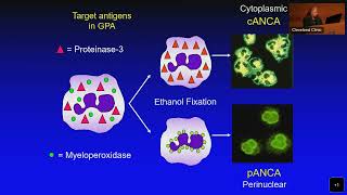
- Eosinophilic granulomatosis with polyangiitis (EGPA) (Churg-Strauss syndrome): 32:04
- Granuloma faciale: 36:45
- Erythema elevatum diutinum (EED): 42:00
This video is geared towards medical students, pathology or dermatology residents, or practicing pathologists or dermatologists. Of course, this video is for educational purposes only and is not formal medical advice or consultation.
Presented by Jerad M. Gardner, MD. Please subscribe to my channel to be notified of new pathology teaching videos.
Follow me on:
Snapchat: JMGardnerMD
Twitter: @JMGardnerMD
Instagram: @JMGardnerMD
Facebook: / jmgardnermd