Published On Sep 25, 2019
📌 𝐅𝐨𝐥𝐥𝐨𝐰 𝐨𝐧 𝐈𝐧𝐬𝐭𝐚𝐠𝐫𝐚𝐦:- / drgbhanuprakash
📌𝗝𝗼𝗶𝗻 𝗢𝘂𝗿 𝗧𝗲𝗹𝗲𝗴𝗿𝗮𝗺 𝗖𝗵𝗮𝗻𝗻𝗲𝗹 𝗛𝗲𝗿𝗲:- https://t.me/bhanuprakashdr
📌𝗦𝘂𝗯𝘀𝗰𝗿𝗶𝗯𝗲 𝗧𝗼 𝗠𝘆 𝗠𝗮𝗶𝗹𝗶𝗻𝗴 𝗟𝗶𝘀𝘁:- https://linktr.ee/DrGBhanuprakash
Gram staining procedure animation - Principle, Procedure, Interpretation
Principle:
--------------
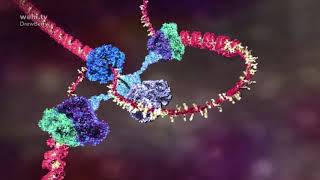
Staining is an auxiliary technique used in microscopic techniques used to enhance the clarity of the microscopic image. Stains and dyes are widely used in the scientific field to highlight the structure of biological specimens, cells, tissues etc.
The most widely used staining procedure in microbiology is the Gram stain, discovered by the Danish scientist and physician Hans Christian Joachim Gram in 1884. Gram staining is a differential staining technique that differentiates bacteria into two groups: gram-positives and gram-negatives. The procedure is based on the ability of microorganisms to retain color of the stains used during the gram stain reaction. Gram-negative bacteria are decolorized by the alcohol, losing the color of the primary stain, purple. Gram-positive bacteria are not decolorized by alcohol and will remain as purple. After decolorization step, a counterstain is used to impart a pink color to the decolorized gram-negative organisms.
Importance of a Gram Stain:
---------------------------------------------
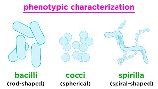
The Gram stain is a very important preliminary step in the initial characterization and classification of bacteria. It is also a key procedure in the identification of bacteria based on staining characteristics, enabling the bacteria to be examined using a light microscope. The bacteria present in an unstained smear are invisible when viewed using a light microscope. Once stained, the morphology and arrangement of the bacteria may be observed as well. Furthermore, it is also an important step in the screening of infectious agents in clinical specimens such as direct smears from a patient.
The Gram stain procedure enables bacteria to retain color of the stains, based on the differences in the chemical and physical properties of the cell wall.
1. Gram positive bacteria: Stain dark purple due to retaining the primary dye called Crystal Violet in the cell wall. Example: Staphylococcus aureus
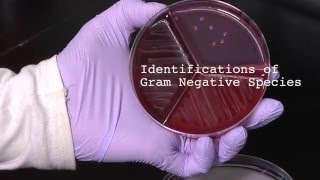
2. Gram negative bacteria: Stain red or pink due to retaining the counter staining dye called Safranin.
Example: Escherichia coli
#gramstainingprocedure #microbiology #usmlevideos #usmle #usmlestep1videos #neetpg #fmge #mbbs #microbiologyvideos #microlectures #animatedmedicalvideos #animatedmicrobiologyvideos #drgbhanuprakash