Published On May 30, 2022
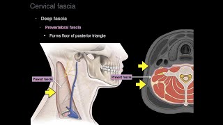
A variant of the deep fascia, in the form of a sheet of pearly-white elastic fibrous tissue that covers a portion of the muscle belly and acts as insertion sites for muscle fibers, while free tendons connect muscles to bones.
Tendons allow the body to move and be flexible while aponeuroses allow the body to be strong and stable. Aponeurosis can thin into a tendon and become a point of origin or insertion for other muscles.
It is also used in pennate muscles, in which the muscle fibers are oriented at an angle to the line of action, typically have two aponeuroses. Muscle fibers connect one to the other, and each aponeurosis thins into a tendon which attaches to bone at the origin or insertion site.
(Comparison to tendon)
• aponeurosis
- - Made of layers of delicate, thin sheaths, primarily of bundles of collagen fibers distributed in regular parallel patterns.
- - Function of binding muscles together or to other tissues and absorbing energy during the movement of the muscle.
- - Hidden under many layers of bones and muscles, so very rare to get injured.
• tendon
- - Tough and rope-like structure.
- - Function of stretching and contracting during muscle movements.
- - Gets injured easily for being located in all the injury-prone areas.
(Examples)
• epicranial aponeurosis (galea aponeurotica): Covers the upper part of the skull.
• erector spinae aponeurosis
• bicipital aponeurosis
• palmar aponeurosis
• posterior intercostal membrane: The internal intercostal muscle is continued to the vertebral column by this thin aponeuroses.
• thoracolumbar fascia
• rectus sheath (rectus fascia)
• aponeurosis of the external oblique
• gluteal aponeurosis
• plantar aponeurosis
Cf. fascia